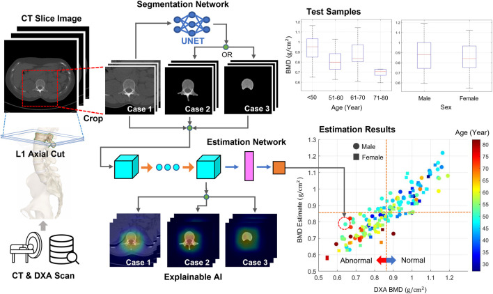
FIGURE 1.
Workflow of this study. From the CT axial images, two image sets were generated. The first set included images cropped around lumbar vertebrae from original images. The second set included images highlighting only the bone body using a segmentation technique. The neural network was trained and tested using either image set for predicting BMDs. DXA BMDs were used as references for supervised learning. Bone diseases, such as osteoporosis and osteopenia, were screened using a T-score converted from every BMD. In addition, the attention map was obtained using XAI techniques to examine the important regions for the BMD prediction.

