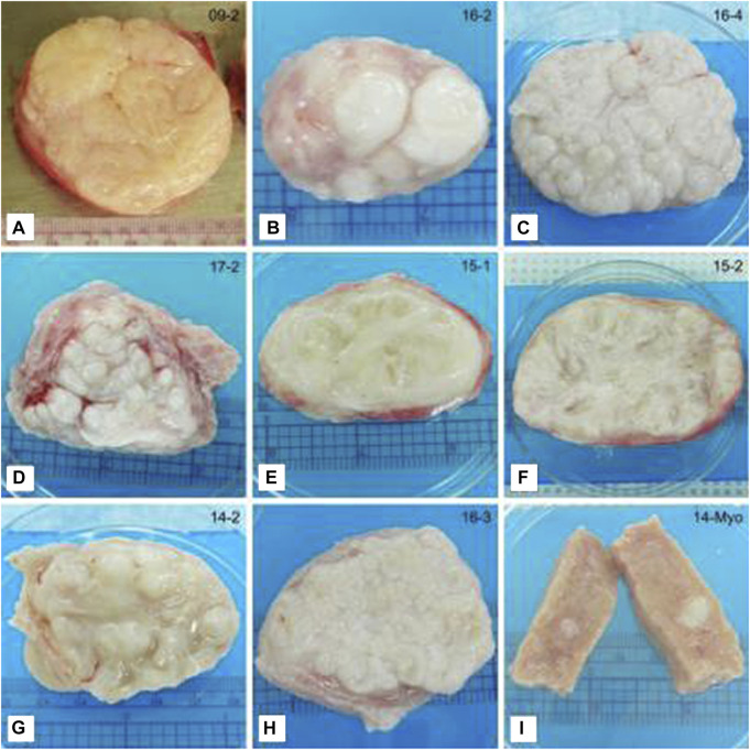
FIGURE 4.
Representative photographs of tissue slices showing differences in the gross appearance of fibroids. (A) Classical irregular whorled pattern. (B–D) Patterns of nodules. (E,F) Trabecular structures. (G) Characteristics of multiple patterns. This example shows a trabecular/nodular pattern. (H) Not categorized. This example shows a tightly gyrated pattern. (I) Myometrial tissue shown for comparison. Note the seedling fibroid embedded in the tissue (white). Ruler (cm) shown for size. This figure and description were adapted from Jayes et al., 2019.

