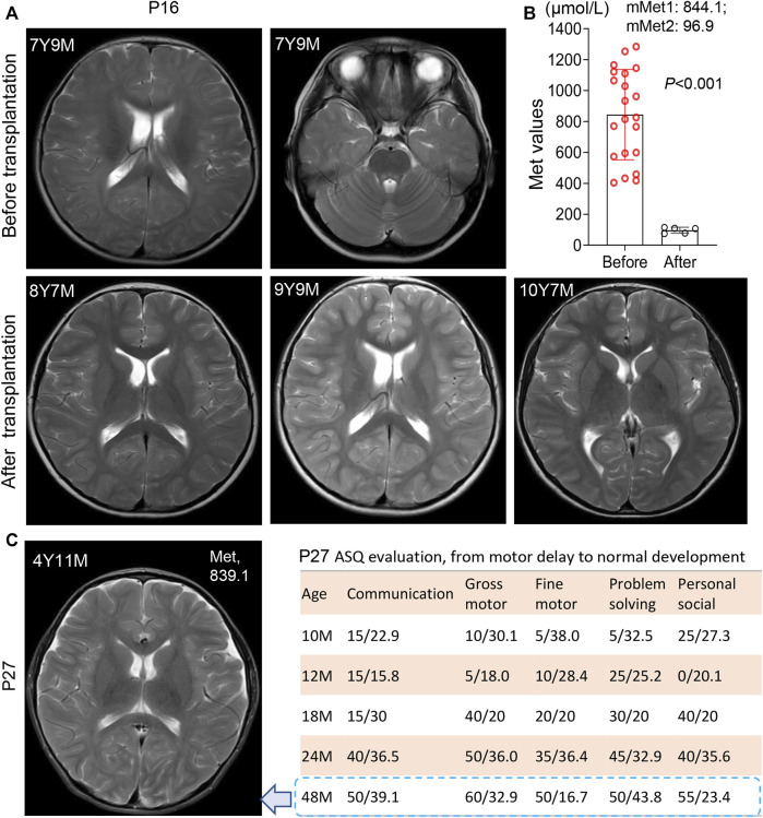
FIGURE 2.
White matter lesions and development status in patient 16 and patient 27. (A) Brain MRI scanning before and after liver transplantation in patient 16 at different time points. 7Y9M = patient at 7 years-9 months old. Patient 16 harbored a heterozygosis c.695 C>T/p. P232L. White matter lesions was reversed after liver transplantation. (B) Significant decrease of Met levels after transplantation in patient 16, from 844.1 μmol/L (before) to 96.9 µmol/L (after). mMet1:844.1 = mean Met value before transplantation was 844.1 μmol/L; mMet2:96.9 = mean Met value after transplantation was 96.9 μmol/L (C) Brain MRI scanning of patient 27 at the age of 4 years 11 m with anomalies in white matter. The plasma Met concentration was maintained at 839.1 μmol/L left panel display his development status measured by ASQ (Ages-Stages Questionnaire). P27 had a normal development after SAMe supplementation. *, p < .05; **, p < .01; ***, p < .001.

