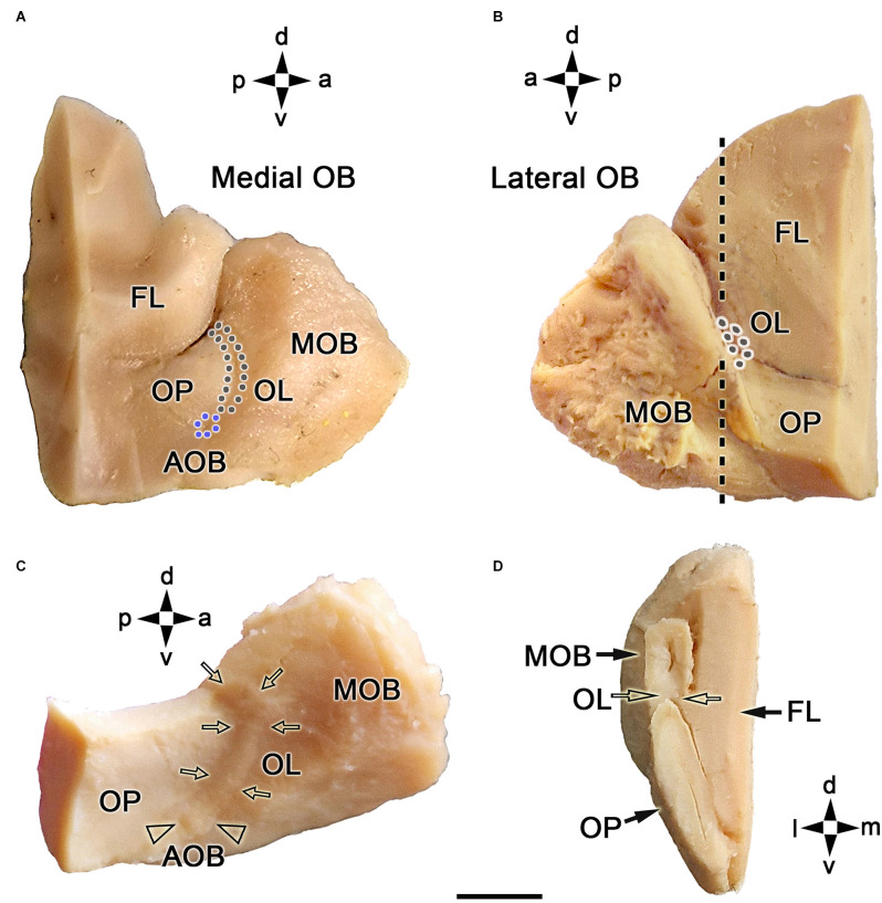
Figure 1.
Macroscopic topographical anatomy of the fox olfactory limbus (OL). (A) Medial view of the left main olfactory bulb (MOB). The olfactory peduncle (OP) and the caudomedial margin of the olfactory bulb are visualized. The accessory olfactory bulb (AOB, blue dots) and the olfactory limbus (OL, black dots) are located along the MOB’s caudal surface. (B) Lateral view of the left main olfactory bulb. The lateral most portion of the OL is located in the caudal margin of the MOB. (C) Dorsolateral view of the right MOB after removal of the frontal lobe (FL). The entire length of the OL (arrows) and the AOB (arrowheads) can be visualized. (D) Caudal view of a transverse section of the left MOB and OP at the level depicted by a dashed line in (B). The OL is located in the caudoventral portion of the MOB (open arrows). a, anterior; d, dorsal; l, lateral; m, medial; p, posterior; v, ventral. Scale bar: 500 μm.

