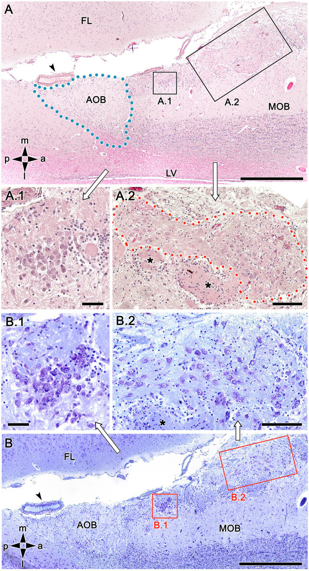
Figure 3.
Histological study of the fox olfactory limbus (OL). (A) General view of the OL, in the horizontal plane. The accessory olfactory bulb (AOB), close to a large artery (arrowhead), is framed by dots. The two most strikingly discernible features of the OL are framed and shown at higher magnification in (A.1) and (A.2): the dense neuronal cluster (A.1) and the macroglomerular complex (MGC, delimited by red dots in A.2), a broad nervous formation consisting of neuronal somata distributed within a neuropil with clearly distinct boundaries. Small, irregularly shaped glomeruli were observed deep to both structures (asterisks). (B) Consecutive, Nissl-stained serial sections showing the morphology of the neuronal somata. Both the denser aggregates (B.1) and the MGC (B.2), possess polygonal, ellipsoidal, and rounded somata. FL, frontal lobe; LV, lateral ventricle; MOB, main olfactory bulb. Orientation: m, medial; l, lateral; p, posterior; a, anterior. Scale bars: (A,B): 500 μm. (A.1,B.1): 100 μm. (A.2,B.2): 250 μm.

