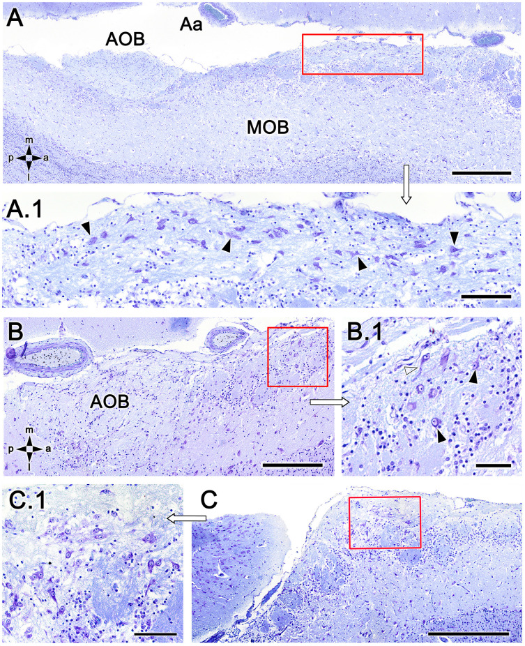
Figure 5.
Nissl-stained sections of the fox olfactory limbus (OL). (A) A ventral section shows the topography of the nervous formation, located on the bulbar surface, superficial to the glomeruli. (A.1) A higher magnification of the red box in (A) shows numerous neuronal somata (arrowheads). (B) Horizontal section at the level of the AOB. Anterior to the AOB, a superficial neuronal cluster surrounded by atypical glomeruli can be observed (red box, magnified in B.1). (B.1) The neuronal somata have oval shapes (arrowheads), and the origin of thick processes is visible (open arrowhead). (C) In this specimen, the neuronal cluster is located in a more anterior position, very close to the pial surface. (C.1) At a higher magnification, the neurons show similar morphology to that of the specimen shown in (B). Aa, artery; AOB, accessory olfactory bulb; MOB, main olfactory bulb. Orientation: m, medial; l, lateral; p, posterior; a, anterior. Scale bars: (A,C): 500 μm. (B): 250 μm. (A.1,C.1): 100 μm. (B.1): 50 μm.

