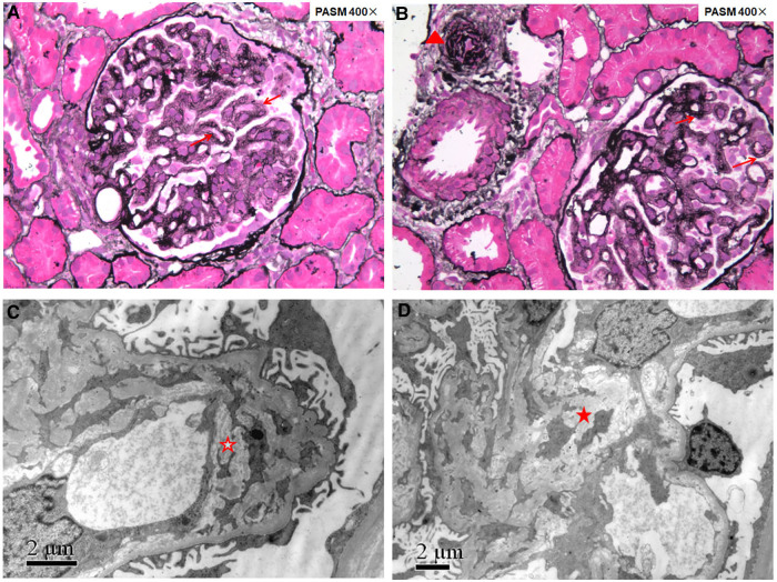
Figure 1.
Renal pathology of patient 1 showing renal thrombotic microangiopathy. Fourteen glomeruli were sampled for light microscopy evaluation (A,B): the glomerular basement membrane thickened diffusely with double contours (red arrows); arterioles showed narrowed lumens with endothelial swelling and intimal onion-skin change (red triangle). Electron microscopy evaluation (C,D): the glomerular basement membrane thickened with subendothelial widening filled with electron-lucent fluffy material, diffusely mesangial interposition along the inner layer of the glomerular basement membrane to form a double contour (red hollow star), and electron transparent mesangial lysis can be seen in the mesangial region (red solid star).

