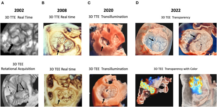
Figure 1.
Examples of technical evolution of 3D echocardiography. In (A) examples are reported of P2 prolapse by RT 3D TTE (top panel); anterior and posterior leaflet prolapse (arrows) by rotational 3D TEE (bottom panel). In (B) examples are reported of P2 flail by RT 3D TTE (top panel), and P1 flail by 3D RT TEE (bottom panel). In (C) an example is reported of TI rendering: P2 prolapse by 3D TTE (top panel) and by 3D TEE (bottom panel). In (D) an example is reported of the “transparency” effect: P2 flail with a detailed visualization of the ruptured chorda by 3D TTE (top panel); mitral regurgitation color Doppler superimposed to the 3D TEE image (bottom panel). 3D, three-dimensional; RT, real-time. TEE, transesophageal echocardiography; TI, transillumination. TTE, transthoracic echocardiography.

