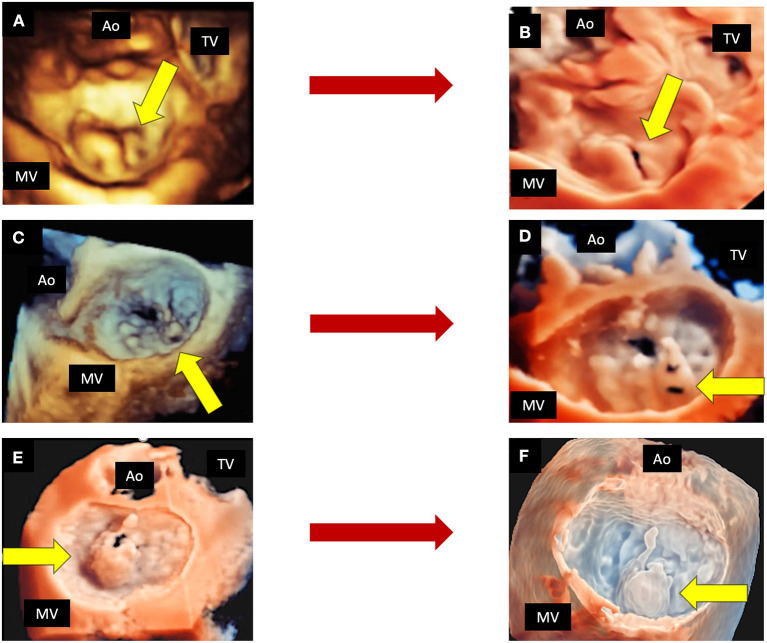
Figure 4.
The role of new 3D tools in echocardiography. Shown are three examples of MVP, in which new tools may improve the quality of imaging. Top panels show a P2 prolapse by standard 3D TTE (A) and by TI rendering (B). Mid panels show a complex P2 prolapse by standard 3D TEE (C) and by TI rendering, which clearly shows the fragile texture of the leaflet (D). Bottom panels show a P2 flail by 3D TEE applying the TI tool (E) and the transparency effect (F). 3D, three-dimensional; Ao, aorta; MV, mitral valve; TEE, transesophageal echocardiography; TI, transillumination. TTE, transthoracic echocardiography; TV, tricuspid valve.

