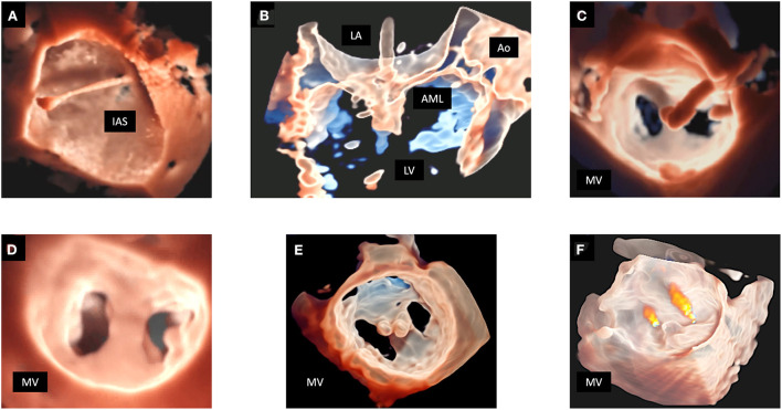
Figure 7.
MitraClip procedure monitoring in a patient with MV prolapse. In (A) TI allows a detailed depiction of the trajectory across the interatrial septum into the left atrium. In (B) the “transparency” effect clearly shows the capture of the MV leaflets by the clip. In (C) TI rendering enhances the visualization the clip before deciding for its release. The lower panels show the result of the procedure. 3D MV reconstruction is displayed by TI rendering from the LA perspective (D), and from the LV perspective with the application of the “transparency” effect, clearly showing 2 clips implanted in the correct position (E). In (F) the residual regurgitant jets are displayed in a 3D reconstruction of the MV with the “transparency” effect and superimposed color Doppler. 3D, three-dimensional; AML, anterior mitral leaflet; Ao, aorta; IAS, interatrial septum; LA, left atrium; LV, left ventricle; MV, mitral valve; TEE, transesophageal echocardiography; TI, transillumination. TTE, transthoracic echocardiography.

