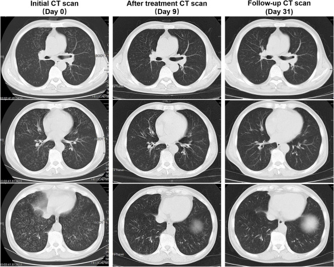
FIGURE 1.
Patient’s computed tomography (CT) scan images at three time points. Initial CT scan (Day 0) showed that bilateral diffuse nodules separated by the fissures and pleura. Some of the nodules have a “tree-in-bud” appearance. After treatment, CT scan (Day 9) showed visible absorption of diffuse nodules in both lungs. Follow-up CT scan (Day 31) showed that the bilateral diffuse nodules absorbed completely.

