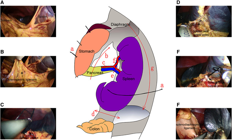
Figure 2.
Operation flow of the SILS. (A) The traction sutures were used to pull the spleen and stomach, such that the splenogastric ligaments were in tension. (B) The splenogastric ligament was cut to expose the pancreatic tail and splenic hilum. (C) The splenic artery was exposed, divided, and ligated. The lower pole (D) and upper pole vein (E) of the spleen was exposed, ligated, and cut off. (F) The splenodiaphragmatic ligament and splenorenal ligament were severed.

