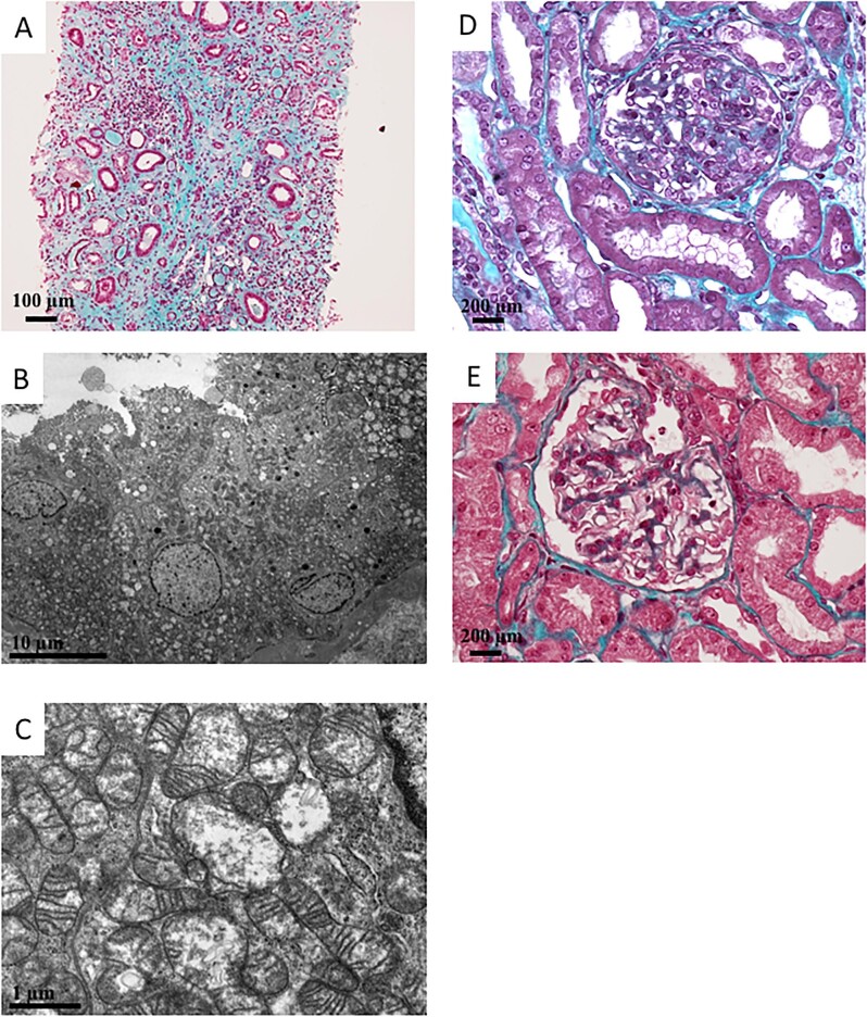
Figure 2:
Histopathological manifestations related to MIDAN. (A) Pathology study by optical microscopy. Magnification ×10, Masson's trichrome staining. Global aspect of severe interstitial fibrosis and tubular atrophy. (B, C) Pathological electron microscopic studies detected abnormal tubular mitochondria that were swollen, with loss of cristae structures. (D) Pathology study by optical microscopy. Magnification ×20, Masson's trichrome staining. Glomerulus presented segmental lesions (e.g. affecting only a portion of the glomerular tuft) that associated proliferation and hypertrophy of podocytes and synechiae between the Bowman's capsule and the tuft. These lesions are compatible with FSGS. (E) Pathology study by optical microscopy. Magnification ×20, Masson's trichrome staining. Glomerulus presented with hypertrophied podocyte lesions (black arrows), but without FSGS lesion.

