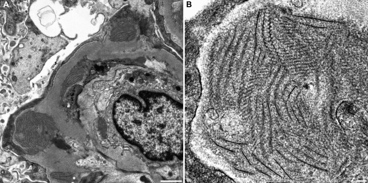
FIGURE 1:
(A) Electron-dense deposits were observed in the subepithelial, intramembranous and subendothelial areas of the GBM under transmission electron microscopy. Extensive foot process effacement of the podocytes and mesangial interposition were also observed. Bar = 1 µm. (B) The deposits at high magnification revealed fibrillary structures with a width of 8–14 nm associated with ladder formation with a periodicity of 25–30 nm. Bar = 100 nm.

