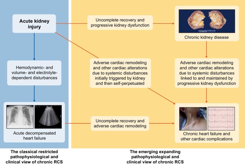
FIGURE 2:
Schematic view of the two pathophysiological and clinical views of acute renocardiac syndrome (RCS) or type 3 cardiorenal syndrome: the classical restricted one (left part of the figure) and the emerging expanding one (right part of the figure). The depicted photographs are the following: chronic kidney disease: macroscopic appearance of end-stage kidney disease. Acute decompensated kidney failure: chest X-ray image of pulmonary oedema (left) and lung ultrasound image of B-lines (right). Chronic heart failure and other cardiac complications: clinical aspect of jugular engorgement (left) and electrocardiographic image of acute myocardial infarction (right).

