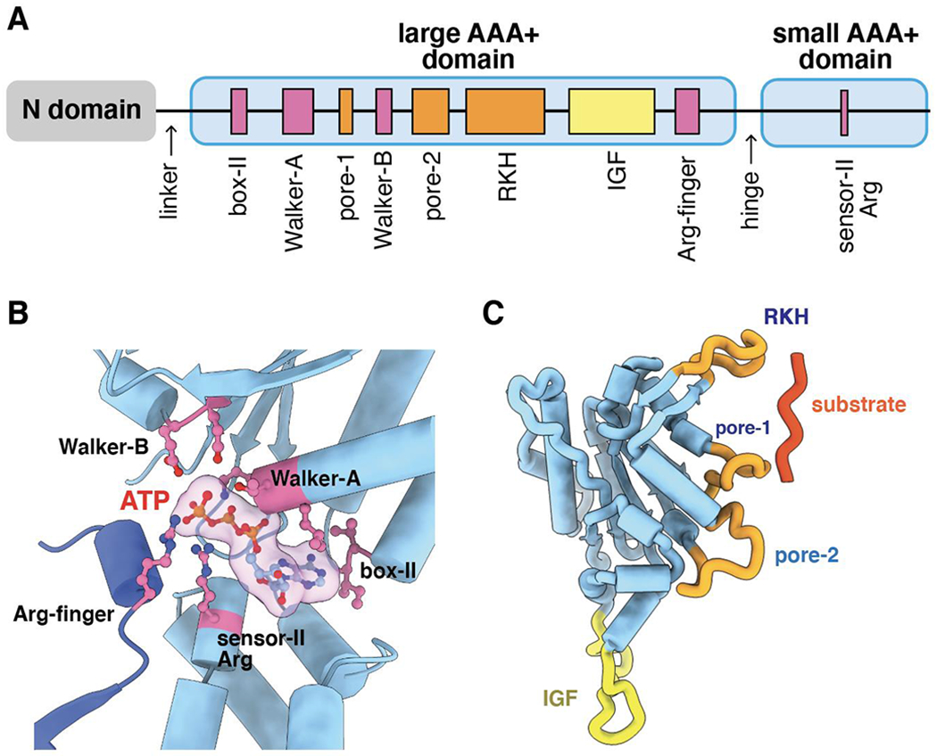
Figure 2.

(A) Cartoon depiction of the domain structure and important sequence motifs in a ClpX subunit. (B) ATP (transparent surface) is contacted by box-II, Walker-A, Walker-B, and sensor-II residues from one subunit (light blue) and by the arginine finger of the neighboring subunit (darker blue). (C) Substrate and ClpP binding loops in a large AAA+ domain of a ClpX subunit.
