
Figure 7.
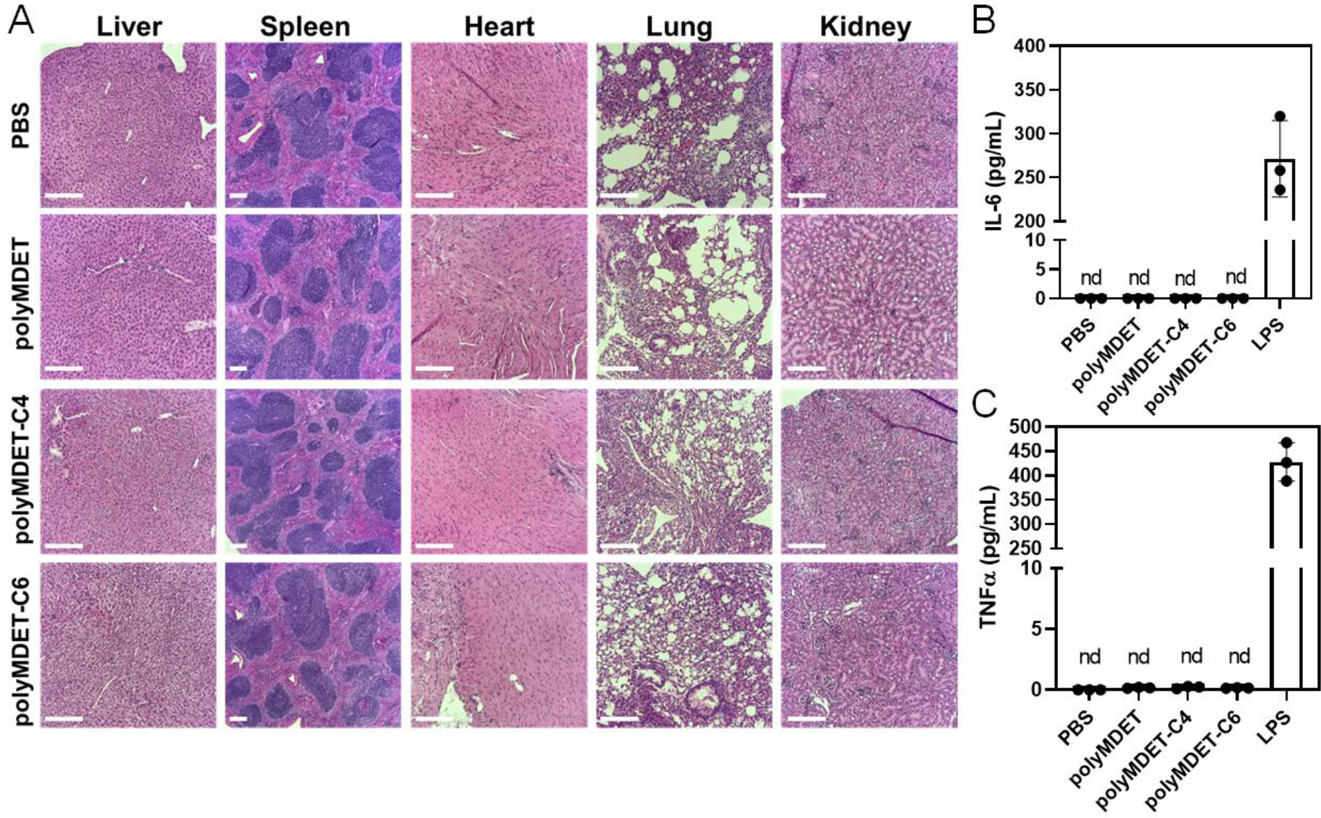
In vivo histological examination and proinflammatory cytokine levels of polyplex-treated mice. (A) Histopathological changes of various organ tissues after single dose intravenous administration of polyplexes (10 μg mRNA) in mice. Scale bar 200 μm. (B) Expression of plasma proinflammatory cytokines after intravenous administration of the polyplexes using ELISA. mRNA polyplexes did not show any proinflammatory cytokine IL-6 and TNFα expression after 24 h. Lipopolysaccharide (LPS) used as positive control induced higher amount of IL-6 and TNFα expression. nd – not detected. N=3 mice per experimental group.