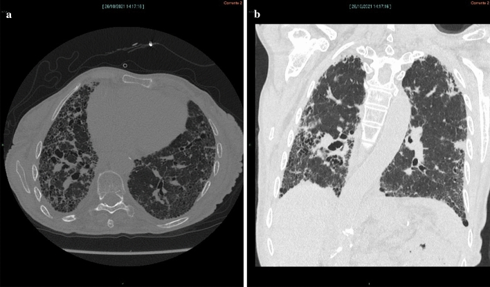
Fig. 1.
Axial computed tomography (CT) image (a) shows diffuse irregular interstitial thickening with extensive honeycombing, more evident in the subpleural region of the right lower lobe; there is evidence of bronchiectasis,irregular pleural and fissural surfaces; and reduced volume of the right lung. Coronal CT reconstruction (b) shows that fibrotic changes are more widespread in the subpleural region of the lower lobes, especially in the right lung with reduced right-lung volume

