Figure 5.
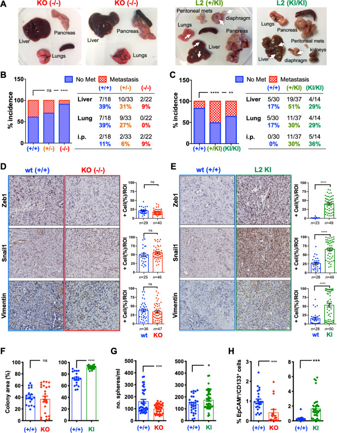
Loxl2 loss and overexpression affects PDAC metastasis and cancer stem cells properties. (A) PDAC tumours and metastases from indicated genotypes (white arrows, metastases). (B–C) Percent incidence of metastasis in (B) wild type (+/+), Het (KPC; Loxl2 +/-) and KO KPC (KPC; Loxl2 -/-) mice or (C) KPC wild type (+/+), KPC; R26L2+/KI and KPC; R26L2KI/KI mice at 17–18 weeks post birth. (**P<0.01, ****p<0.0001; ns, not significant; contingency analysis, two-sided Fisher’s exact test). (D–E) Left: Representative immunohistochemical stainings for indicated proteins in tumour sections from (D) wild type (+/+) KPC and KO KPC (KPC; Loxl2-/- ) mice or (E) wild type (+/+) KPC and KPCL2KI (KPC; R26L2KI/KI) mice. Scale bar=250 µm. Right: Quantification of per cent positive (+) cells/region of interest (ROI). (****P<0.0001; ns, not significant; unpaired Student’s t-test). (F) Mean number % colony area ±SEM, determined 11 days post seeding in indicated cell lines (n=3 cell lines/genotype, ****p<0.0001, ns, not significant, two-sided t-test with Mann-Whitney U test). (G) Mean number (no.) spheres/mL ±SEM, 7 days post seeding, in tumour cell lines from indicated genotypes (*p<0.05, ***p<0.001, unpaired Student’s t-test). (H) Mean percentage of EpCAM+/CD133+ cells ±SEM, in tumour cell lines from indicated genotypes (***p<0.001, two-sided t-test with Mann-Whitney U test). Het, heterozygous; KI, knock-in; KO, knockout; Loxl2, lysyl oxidase-like protein 2; PDAC, pancreatic ductal adenocarcinoma; wt, wild type.

