Figure 1.
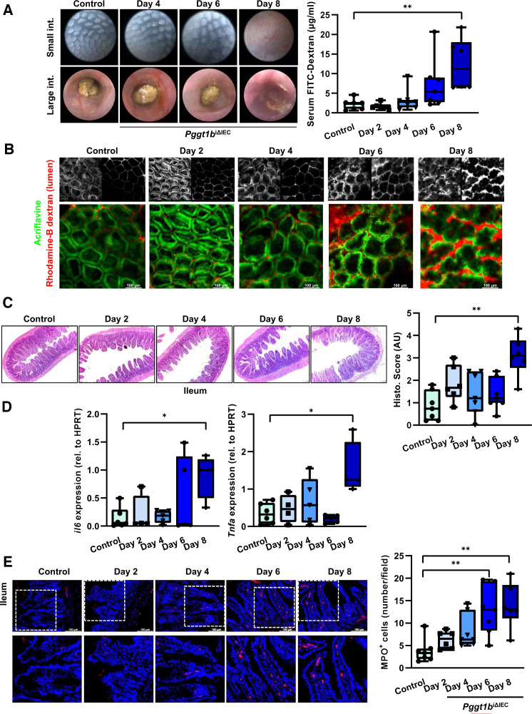
Intestinal disease due to inhibition of prenylation within IECs over time. (A) Assessment of tissue integrity in vivo/ ex vivo and intestinal permeability in vivo. Endoscopy pictures of colon and small intestine tissue (left); transmucosal passage of orally administered FITC-Dextran (4 KDa); serum concentration (µg/mL) (right). (B) Representative pictures of intravital microscopy analysis of barrier function of small intestine using luminal acriflavine (green) and rhodamineB-dextran (red). (Control, n=8; day 2, n=6; day 4, n=5; day 6, n=5; day 8, n=2). (C) Histology analysis of ileum tissue using H&E staining. Representative pictures (left), and corresponding score (right). (D) Gene expression of IL-6 and TNF-α in ileum tissue (RT-qPCR; six independent experiments). (E) MPO immunofluorescence staining in cross-sections from ileum. Representative pictures (left), and corresponding quantification (right). Data are expressed as box-plots (Min to Max); seven independent experiments, except where indicated. One-way ANOVA, Dunnett’s multiple comparisons test. *P≤0.050; ** P≤0.001. ANOVA, analysis of variance.

