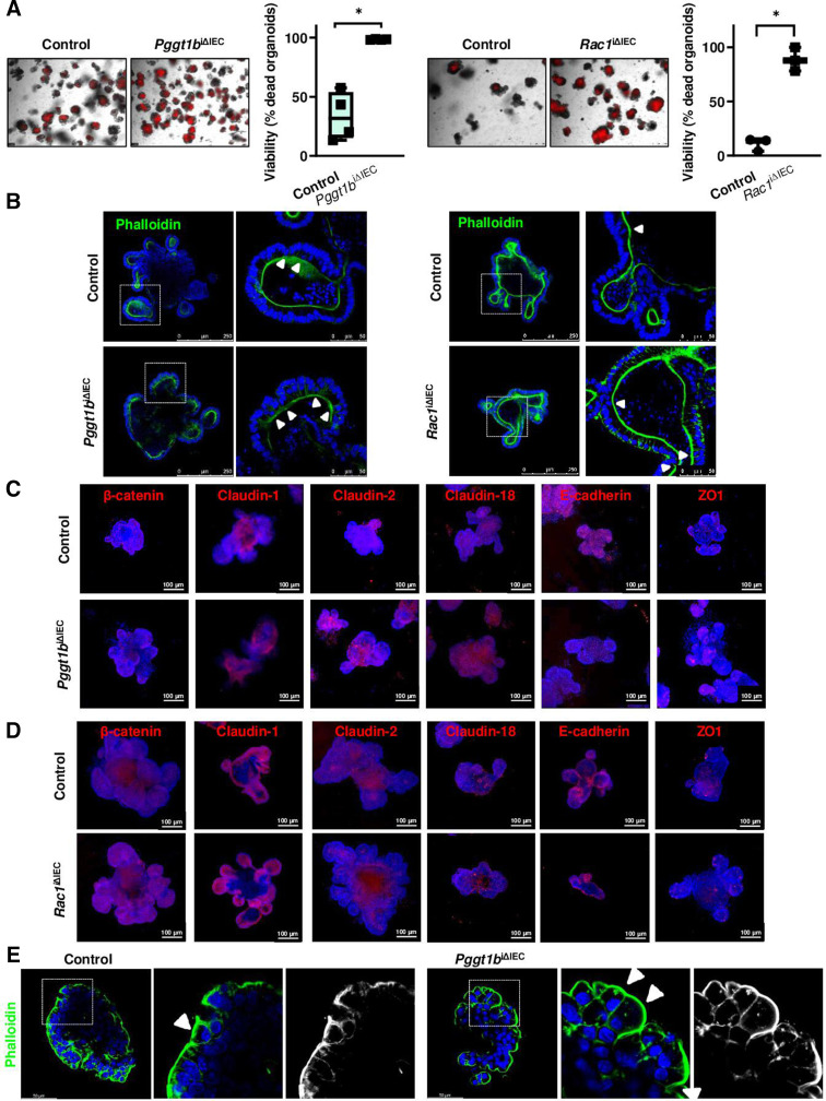
Figure 8.
Epithelial intrinsic mechanisms in small intestine organoids. GGTase-deficient and RAC1-deficient organoids generated from small intestinal crypts. (A) Cell viability measured by PI incorporation (red). Representative pictures (left), and corresponding quantification (% of dead organoids) (right). (Pggt1b, four experiments; Rac1, 3 experiments). (B) F-actin fibre staining using AlexaFluor488-phalloidin (green) (Pggt1b, five experiments; Rac1, four experiments). White arrows indicate funnel-like structures or arrested cell shedding events. (C, D). Analysis of candidate AJC proteins by immunostaining. Maximum projection from z-stacks (system optimised). (C) GGTase-deficient organoids. Four experiments; except for β-catenin and Claudin-1, three experiments. (D) RAC1-deficient organoids. Three experiments; except for claudin-2, five experiments. (E) Apical-out GGTase-deficient organoids, representative pictures of Phalloidin staining. One experiment. Data are expressed as box-plots (Min to Max). Paired t-test. *P≤0.050. AJC, apical junction complex.

