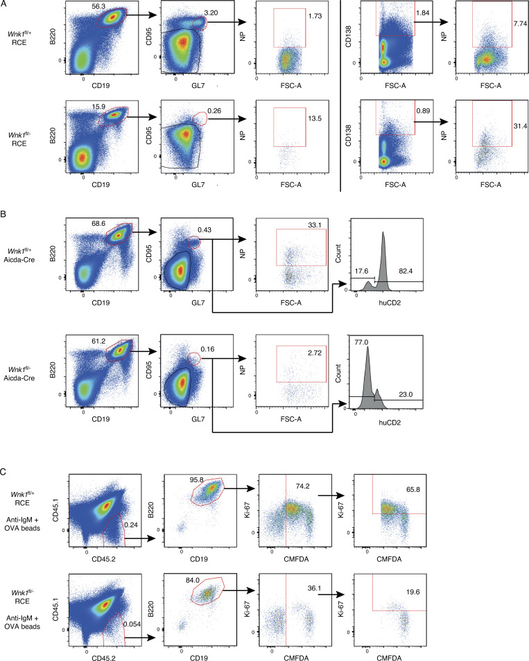
Figure S5.
WNK1-deficient B cells fail to generate antigen-specific GCB cells and plasma cells and to divide in vivo. (A) Flow cytometric analysis of splenocytes from mice immunized with NP-CGG in alum treated as described in Fig. 6 A, showing gating strategy for NP-specific germinal center cells (B220+CD19+CD95+GL7+NP+) and NP-specific plasma cells (CD138hiNP+) for control (top row) and mutant mice (bottom row). Numbers on dot plots indicate percentages of cell populations within gates (red boxes). (B) Flow cytometric analysis of splenocytes from mice immunized with NP-CGG in alum treated as described in Fig. 6 D, showing gating strategy for NP-specific germinal center cells (B220+CD19+CD95+GL7+NP+) and a histogram of human CD2 (huCD2) surface expression on GCB cells (B220+CD19+CD95+GL7+) for control (top row) and mutant mice (bottom row). Numbers in dot plots indicate percentages of cell populations within gates (red boxes), numbers on histogram indicate percentage of GCB cells that are negative or positive for huCD2 expression. (C) Flow cytometric analysis of splenocytes from mice treated as described in Fig. 7 A, showing gating strategy for control (top row) and mutant (bottom row) B cells that had been pre-treated with beads conjugated with both anti-IgM and OVA (CD45.1−CD45.2+B220+CD19+CMFDA+) and transferred into a CD45.1+CD45.2+ host, showing analysis of % transferred B cells that were Ki67+. Numbers on dot plots indicate percentages of cell populations within gates (red boxes).

