Fig. 1.
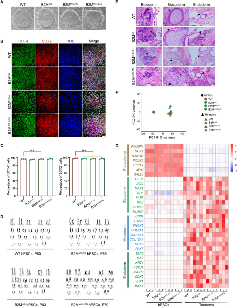
Hypoimmunogenic human pluripotent stem cells (hPSCs) retain normal self-renewal and multilineage differentiation. A Bright-field images of wild-type (WT) and hypoimmunogenic hPSCs cultured more than 30 passages. Scale bar, 100 µm. B Immunostaining of OCT4 and SOX2 in WT and hypoimmunogenic hPSCs. Nuclei were stained with Hoechst (HOE). Scale bar, 75 µm. C FACS quantification showing the percentage of OCT4+ and SOX2+ cells of WT and hypoimmunogenic hPSCs. Data are presented as mean ± SEM. n.s., no significance, n = 3, Student’s t test. D Karyotype analysis of the WT and hypoimmunogenic hPSCs. E H&E staining identified three germ layers in teratomas from WT and hypoimmunogenic hPSCs. Typical tissues were marked by arrowheads (left panel, neural tube-like tissues for ectoderm; middle panel, cartilage-like tissues for mesoderm; right panel, intestine-like tissues for endoderm). Scale bar, 100 μm. F Principal component analysis (PCA) plot of WT, hypoimmunogenic hPSCs and teratomas formed by WT and hypoimmunogenic hPSCs. G Heatmap of differentially expressed signature genes in RNA-seq data of pluripotency, ectoderm, mesoderm and endoderm in WT, hypoimmunogenic hPSCs and their teratomas

