. 2022 Oct 16;21(1):e130762. doi: 10.5812/ijpr-130762

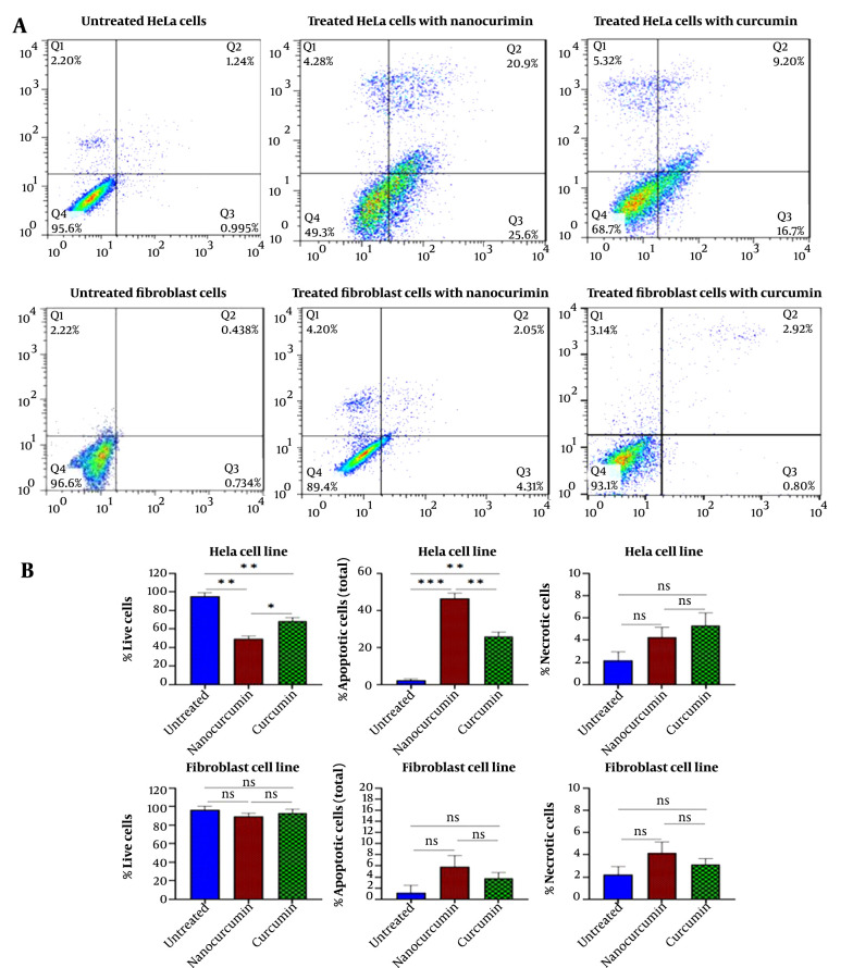
Figure 2. Flow cytometric diagram obtained from annexin/PI test to evaluate cellular apoptosis: A, Apoptosis assay results for cancerous HeLa cells after treatment with 15 μm of nano-curcumin and 50 μm of curcumin. Apoptosis assay results for fibroblast cells in the same condition. B, 46.5% of HeLa cells treated with 15 μM nano-curcumin were destroyed through apoptosis. Free curcumin at a higher concentration (50 μM) could induce apoptosis in 25.9% of cells. The apoptosis test for fibroblast cells showed that nano-curcumin (15 μM) and curcumin (50 μM) had no significant effect on cell apoptosis of normal cells. (Significant at * P < 0.05, ** P < 0.01, *** P < 0.001).

Figure 2.