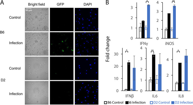
FIG 6.
Experimental validation of macrophages and proinflammatory cytokines after infection by macrophage phagocytosis assay and qRT-PCR. (A) Macrophage phagocytosis assay revealing better clearance of pathogens by macrophages from D2 mice than those from B6 ones. Macrophages were differentiated from the bone marrow cells. Fluorescent images were obtained with a Leica DMi8 Thunder fluorescence microscope (Leica Camera, Wetzlar, Germany) at 550-nm excitation for green fluorescent protein (GFP) expressed in S. Typhimurium strain 14028GFP and 395-nm excitation for DAPI. (B) qRT-PCR analysis for expression of five proinflammatory cytokines (IFN-γ, iNOS, IFN-β, IL-6, and IL-8) after S. Typhimurium infection between B6 and D2 strains by qRT-PCR (n = 2 for each group). Error bars represent mean ± standard deviation (SD). *, P < 0.05; **, P < 0.01.

