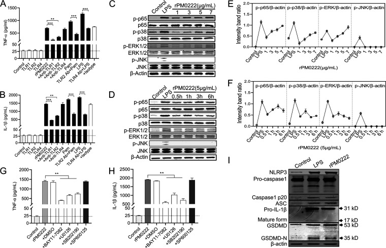
FIG 2.
Secretion of TNF-α and IL-1β in response to rPM0222 is dependent on TLR1/2 and NF-κB/MAPK signaling. (A and B) The cells were pretreated with 5 μg/mL of anti-TLR1, -TLR2, and -TLR4 or an IgG isotype-matched control antibody for 1 h, followed by stimulation with rPM0222 (5 μg/mL) for 6 h. The cell culture supernatants were collected, and the levels of TNF-α and IL-1β were measured using ELISA. Pam, Pam3CSK4. (C and D) The murine peritoneal exudate macrophages were incubated with rPM0222 (1, 3, 5, or 7 μg/mL) for 6 h or incubated with rPM0222 (5 μg/mL) for the indicated times (0.5, 1, 3, or 6 h). Cell lysates were prepared, and the phosphorylation/activation status of p65, ERK1/2, p38, and JNK was analyzed using Western blotting. (E and F) The murine peritoneal exudate macrophages were incubated with rPM0222 (1, 3, 5, or 7 μg/mL) for 6 h or 5 μg/mL rPM0222 for the indicated times (0.5, 1, 3, or 6 h). The intensity band ratio of activation status of p65, ERK1/2, p38, and JNK to that of β-actin was analyzed. (G and H) The murine peritoneal exudate macrophages were pretreated with U0126 (+U0126; 20 μM), SB203580 (+SB202190; 20 μM), SP600125 (+SP600125; 20 μM), BAY11-7082 (+BAY11-7082; 20 μM), or dimethyl sulfoxide (0.01%) (+DMSO) for 1 h, followed by stimulation with rPM0222 (5 μg/mL) for 6 h. Further, the TNF-α and IL-1β levels in cell culture supernatants were measured using ELISA. (I) The murine peritoneal exudate macrophages were incubated with 5 μg/mL rPM0222 for 6 h. Cell lysates were prepared, and the levels of NLRP3, ASC, procaspase-1, p20, mature IL-1β, GSDMD, and GSDMD-N were measured using Western blot analysis. Data are expressed as the mean ± standard deviation from three independent experiments. **, P < 0.01; ***, P < 0.001. The experiments were performed at least three times.

