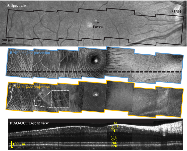
Fig. 6.
Representative wide-field inner retina images from subject S1 collected with the FDA FDML AO system. A. Spectralis macular scan. En face projection views over a -14° to 14° eccentricity range from seven 4.5°×4.5° FOV AO-OCT scans of B. NFBs at NFL and C. macrophages just above the ILM. Inset shows magnified view of one representative macrophage cell at 8° eccentricity. D. AO-OCT B-scan view corresponds to the black dashed line in B.

