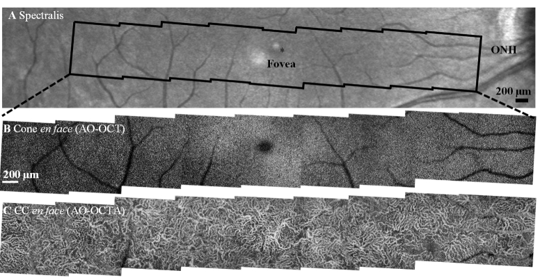
Fig. 10.
Representative wide-field images of outer retinal features from subject S1 collected with the FDA FDML AO system. A. Spectralis macular scan. En face projection views over -11° to 11° eccentricity range from nine 3°×3° FOV scans. B. AO-OCT intensity projection (IS/OS to COST) shows better delineation of the cone photoreceptor mosaic compared to AO-SLO montage from the same region (Fig. 7) due to the exclusion of signals from neighboring rod photoreceptors and underlying RPE cells. C. Corresponding AO-OCTA scans of the choriocapillaris.

