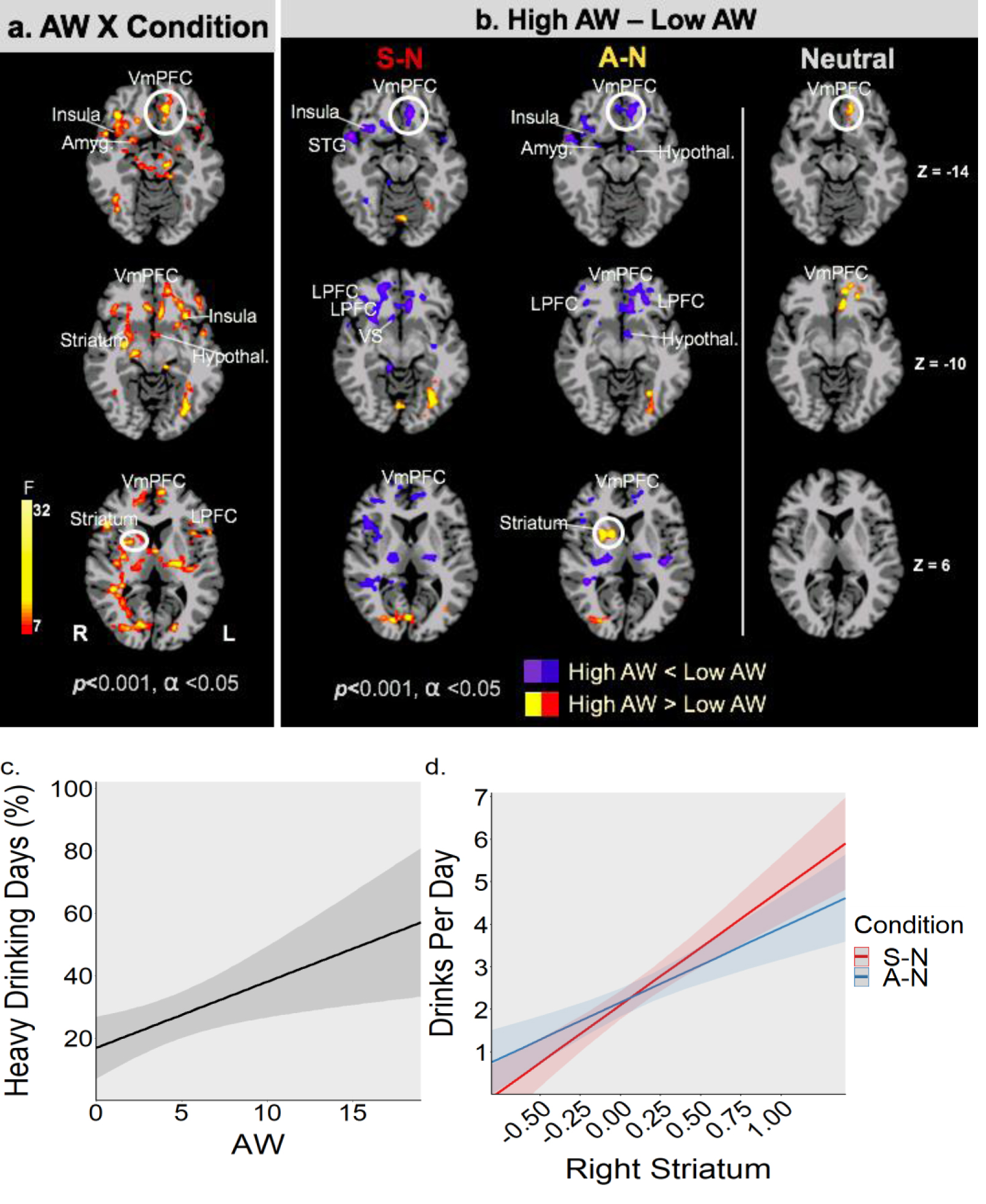
Figure 3.

a: Significant whole brain voxel-wise LME analysis indicates AW (continuous scores) X Condition (Stress-S, alcohol cue-A, neutral-N) interaction effects (shown in red/yellow) in functional brain responses in the VmPFC, dorsolateral PFC, insula, amygdala, striatal, hypothalamic regions, consistent with brain regions involved in functional regulation of stress and reward cue responses (FWE corrected at p<0.001, cluster corrected at α<.05). Figure 3b: Post-hoc secondary analyses to understand the AW Symptom X Condition significant brain effects shown in 3a was conducted by dividing AUD patients into high and low AW scores (median split). Figure 3b shows disrupted VmPFC and related limbic and striatal responses to functional challenge of stress (S) and alcohol cue (A) relative to neutral (N) and under neutral-relaxed (N) state responses (FWE p<.001, α<.05). As hypothesized, VmPFC and lateral PFC regions as well as limbic (Amyg: amygdala, insula, Hypothal: hypothalamus) and striatal (VS: ventral striatum) show reduced/blunted responses during stress and alcohol cue challenge in high AW versus low AW group (blue/purple shows hypoactivation), while Right Striatum (dorsal) shows hyperactive responses during alcohol cue exposure (red/yellow shows hyperactivation). Furthermore, hyperactive and disrupted VmPFC functioning during exposure to neutral-relaxing images is shown in high AW relative to the low AW group. (see Supplemental Table ST1). Figure 3c: Higher AW (CIWA-Ar continuous scores) at intake prospectively predicted higher 2-week drinking during early treatment (Percent heavy drinking days, p <0.01). Error bands are ± SEM. Figure 3d: Higher R dorsal striatum response during challenge (A: alcohol cue; S: stress) relative to neutral (A-N or S-N) predicted higher number of drinks consumed on a drinking day (S-N: Drinks Per Day; p < 0.01; A-N: Drinks Per Day; p <0.035), in the early treatment phase. Error bands are ± SEM.
