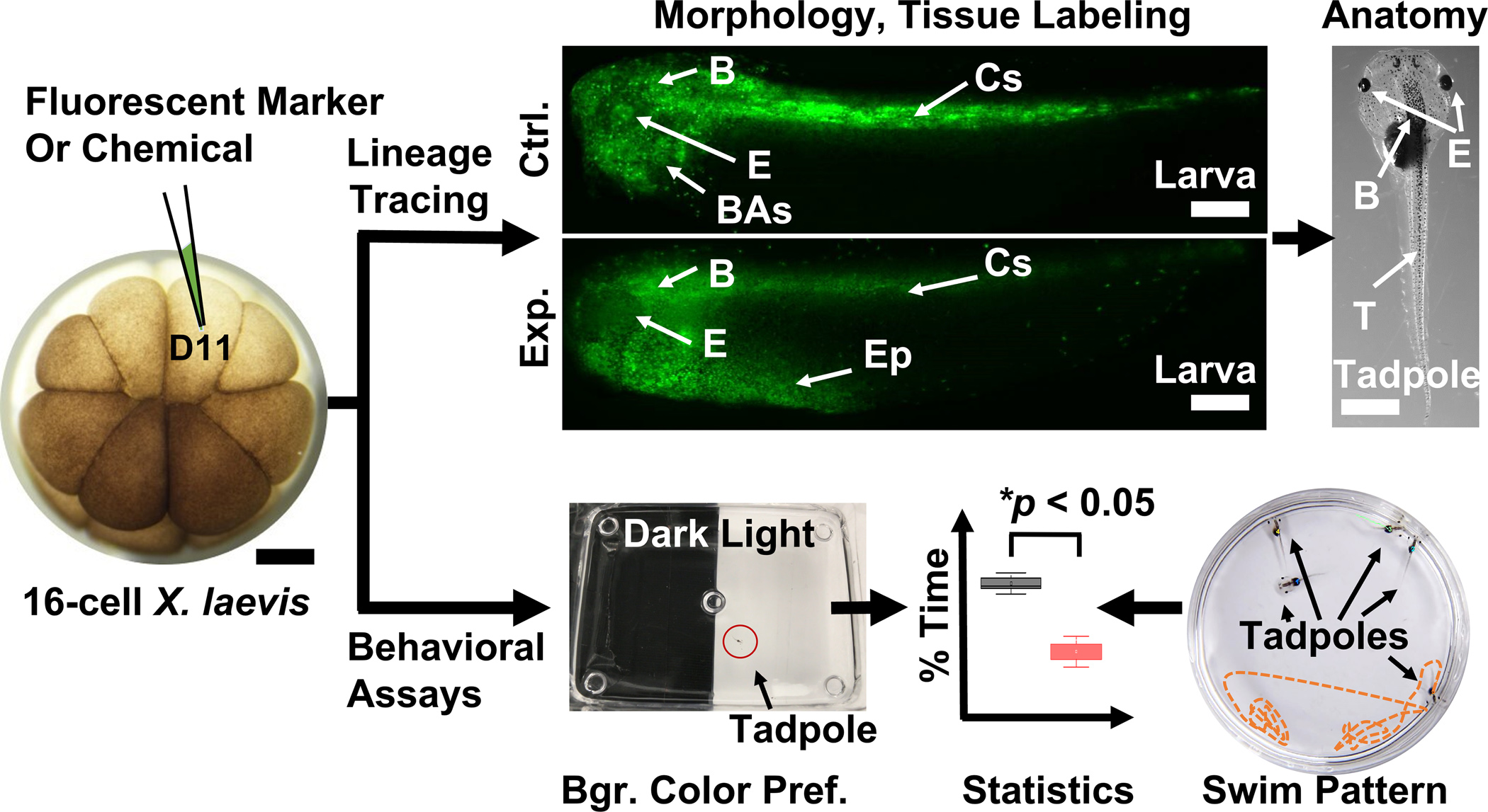
Figure 4.

Techniques to investigate chemistry and function during development. (Top panel) Analysis of cell fate, morphology, and anatomy following fluorescence lineage tracing of the left dorsal-animal midline cell (D11) in control (Ctrl.) and experimental (Exp.) X. laevis larvae. (Bottom panel) Background color preference and swim patter assays evaluating behavior in X. laevis tadpoles. Key: B, brain; BAs, branchial arches; Cc; central somites; E, eye; Ep; epidermis. Scale bars = ~250 μm (embryo, larvae), ~1.5 mm (tadpole). (Adapted with permission from Anal. Chem. 2017, 89, 13, 7069–7076. Copyright 2017 American Chemical Society.)
