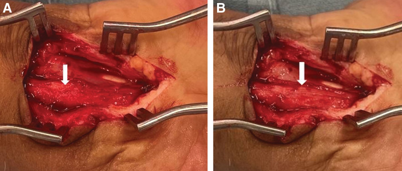
Fig. 4.
Patient B. A, Thick epineurium is seen. B, External neurolysis is performed until the fascicular pattern of the median nerve is visualized, seen here on the ulnar aspect of the nerve, under loupe magnification. In this case, further neurolysis was performed to also decompress the thick, compressive epineurium on the radial aspect of the nerve (not pictured).

