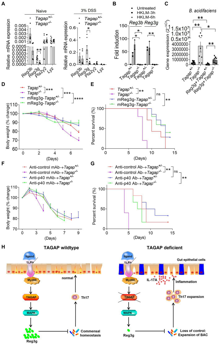
Figure 6.
Reg3g recombinant protein or anti-p40 MAb rescue the severe phenotype of TAGAP-deficient mice in colitis. (A) Colonic tissues were isolated from littermate control mice or TAGAP-deficient mice with or without DSS treatment (day 5 of DSS treatment), followed by real-time PCR analysis of indicated gene expression, n = 7. (B) Colonic tissues isolated from littermate control mice or TAGAP-deficient mice were treated with HKLM for 3 or 6 h, followed by real-time PCR analysis of indicated genes. (C) Littermate control mice or TAGAP-deficient mice were oral gavaged with recombinant reg3g proteins daily for 1 week (5 μg/mouse/day), and feces were collected and analyzed by real time PCR for the indicated bacterial strains, n = 12, 8, 10 and 10. (D,E) Littermate control mice or TAGAP-deficient mice were oral gavaged recombinant reg3g proteins daily for 4 weeks (5 μg/mouse/day), followed by 2.5% DSS treatment for 5 days. Mice weight curve (D) and survival curve (E) were done in separate experiments, n = 7. (F,G) Littermate control mice or TAGAP-deficient mice were intraperitoneal injected anti-control MAb or anti-p40 MAb at day 1, 3, 5 and 7 after DSS treatment (100 μg/100 μl 1 × PBS/mouse). Mice weight curve (F) and survival curve (G) were done in separate experiments, n = 6. (H) Model of TAGAP regulating gut microbiota and Th17 cells abundance in the gut, and affecting IBD susceptibility. *p < 0.05, **p < 0.01, ***p < 0.001, ****p < 0.0001 based on 2-way ANOVA (D,F), Log-rank (Mantel-Cox) Test for panels (E,G) and unpaired T test (A–C). Data are representative of three independent experiments.

