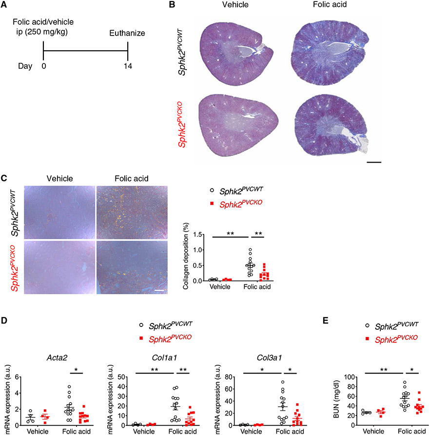
Fig. 2. Sphk2 deletion in renal perivascular cells ameliorates kidney fibrosis in the folic acid mouse model.
(A) Protocol for inducing kidney fibrosis by injecting folic acid (for B to E). Sphk2PVCWT and Sphk2PVCKO mice were given folic acid (250 mg/kg, ip) and euthanized at day 14. (B) Representative Masson’s trichrome staining of collagen in kidney sections at day 14. (C) Picrosirius red staining of kidney (polarized microscopy) with quantification of red/yellow birefringence of mature collagen fibers as a percentage of the total surface area of kidney section at day 14. n = 4 to 13 per group. (D) Acta2, Col1a1, and Col3a1 transcript expression (from whole kidney) at day 14. n = 4 to 13 per group. (E) Blood urea nitrogen (BUN) concentrations at day 14. n = 4 to 13 per group. Scale bars, 1 mm (B) and 200 μm (C). Data are represented as means ± SEM. *P < 0.05 and **P < 0.01; two-way ANOVA followed by post hoc multiple comparisons test (Tukey’s).

