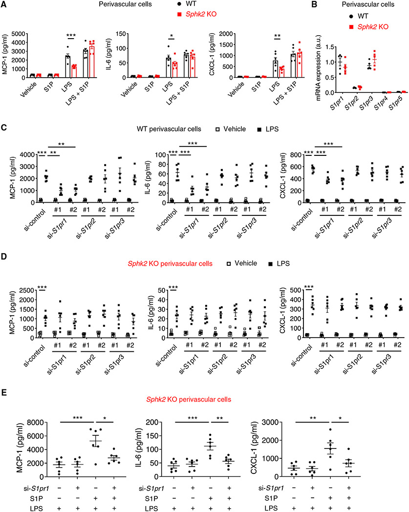
Fig. 4. S1P produced by SphK2 is exported to the extracellular space and binds to S1P1 to enhance inflammatory signaling in perivascular cells.
(A to E) Experiments were performed using primary kidney perivascular cells isolated from Sphk2PVCWT [wild-type (WT)] and Sphk2PVCKO (Sphk2 KO) mice. (A) MCP-1, IL-6, and CXCL-1 concentrations in supernatants of primary kidney perivascular cells treated with LPS (100 ng/ml) for 24 hours with or without S1P supplementation (100 nM). (B) S1pr1-5 transcript expression at baseline. (C and D) MCP-1, IL-6, and CXCL-1 concentrations in supernatants of WT (C) and Sphk2 KO (D) kidney perivascular cells at 24 hours after treatment with LPS (100 ng/ml). Cells were treated with control, S1pr1, S1pr2, or S1pr3 siRNA before LPS stimulation. (E) MCP-1, IL-6, and CXCL-1 concentrations in supernatants of Sphk2 KO kidney perivascular cells treated with LPS (100 ng/ml) for 24 hours with or without S1P supplementation (100 nM). Cells were treated with control or S1pr1 siRNA before LPS stimulation. n = 6 (A, C, D, and E), n = 5 (B) per group. Data are represented as means ± SEM. *P < 0.05, **P < 0.01, and ***P < 0.001 by two-way (A to D) or one-way (E) ANOVA followed by post hoc multiple comparisons test (Tukey’s).

