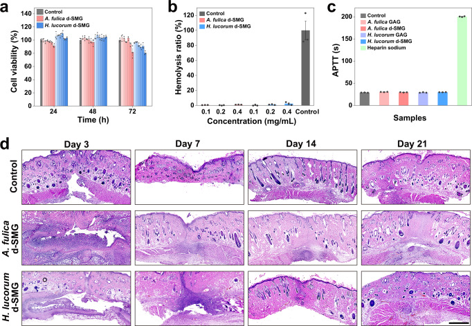
Fig. 3. Biocompatibility and biodegradability of d-SMGs.
a Effect of different concentrations (0.05, 0.1, 0.2, 0.5, 1.0, and 2.0 mg/mL from left to right in the graph) of d-SMG on cell viability by MTT method (n = 3 cells examined over 3 independent experiments in each group). b Effect of d-SMGs on hemolysis (n = 3 biologically independent samples in each group). c Effect of d-SMGs and their s-GAGs on APTT of human coagulation control plasma (n = 3 biologically independent samples in each group). d H&E staining images of skin tissue after subcutaneous embedding of d-SMGs in SD rat skin incision model (n = 3 biologically independent samples in each group). Data are presented as mean values ± SEM. Scale bar, 1 mm (images in d). Source data are provided as a Source data file.

