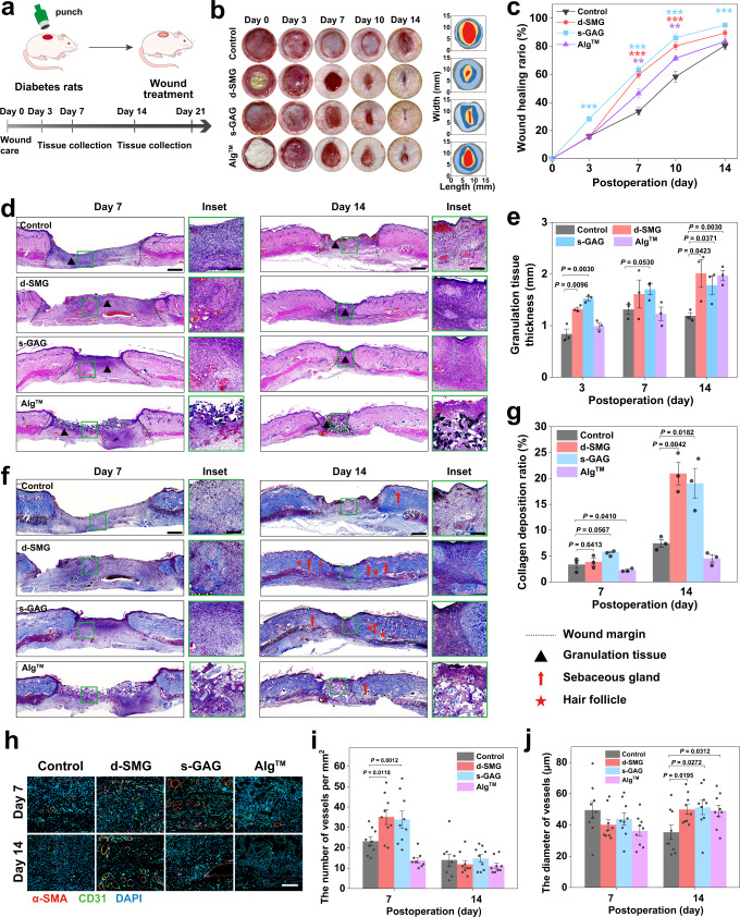
Fig. 6. Effects of A. fulica d-SMG and s-GAG on chronic wound healing in diabetic rats.
a Schematic illustration of the diabetic wound model in SD rats. b Representative images of the wound healing behavior and dynamic wound healing process on day 0, 3, 7, 10, and 14. c The wound healing ratio on day 0, 3, 7, 10, and 14 (n = 7 rats created over 2 independent wounds in each group, two-tailed t test was used, *P < 0.05, **P < 0.01, ***P < 0.001). d H&E-stained images of the wound tissue on day 7 and 14. e The granulation tissue thickness in wound tissue on day 7 and 14 (n = 3 biologically independent samples in each group). f Masson staining of the wound tissue on day 7 and 14. g The collagen deposition ratio of wound tissue on day 7 and 14 (n = 3 biologically independent samples in each group). h Representative images of CD31 and α-SMA immunostaining in wound tissue on day 7 and 14. I, j Quantification of the number (i) and diameter (j) of vessels in the wound tissue (n = 9 biologically independent samples in each group). For (c, e, g, i, and j), two-tailed t test was used. Data are presented as mean values ± SEM. Scale bars, 1 mm (images in d, f), 200 μm (inset in d, f), 100 μm (images in h). Source data are provided as a Source data file.

