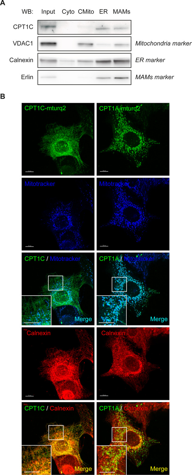
Fig. 4. Human CPT1C is mainly localized in ER and mitochondria–ER contact sites in cancer cells.

A MCF7 cells were seeded, collected and isolated by cellular subfractionation and Percol gradient in order to separate cytoplasm (Cyto), crude mitochondria (CMito, mitochondria and other contaminating membranes), microsomes (fraction containing the ER), and mitochondria-associated membranes (MAMs, fraction containing mitochondria–ER contacts). Also kept was whole lysate (input). All fractions were prepared for the same total protein concentration and processed by Western blotting, and equal volumes of each fraction were loaded. Endogenous CPT1C was mainly localized at ER and MAMs. B MCF7 cells were transfected with human CPT1C or CPT1A tagged with mTurquoise2 (mTurq2); 48 h later, Mitotracker Orange (200 nM) was added, and incubation was 30 min. The cells were then fixed. and calnexin (used as an ER marker) was detected by immunocytochemistry. CPT1C mainly colocalized with the ER (orange spots), while CPT1A colocalized with mitochondria (light blue spots). The colocalization of CPT1C with mitochondria (the few light blue spots) may correspond to ER–mitochondria contacts. The scale bar in B is 10 µm.
