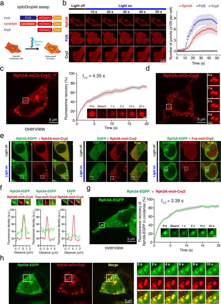
Fig. 1. Rph3A forms condensates with liquid properties in living cells.
a Schematic illustration of the optoDroplet assay in HEK293 cells. Candidate genes were fused with mCherry and Cry2. The RNA-binding protein FUS was chosen as a positive control. Unfused mCherry-Cry2 was used as a negative control. Upon blue light exposure, proteins with phase-separation capacity formed condensates in cells. b OptoDroplet assay of Rph3A performed in HEK293 cells. The number of puncta per cell was counted and plotted. Data are displayed as the mean ± SEM (n = 6 cells per group). c Representative images and quantification of fluorescence recovery from the FRAP analysis of Rph3A droplets. The time point of bleaching was 0 s. The data are displayed as the mean ± SEM (n = 6 droplets). d Fusion events of droplets composed of Rph3A-mCherry-Cry2 in HEK293 cells captured by time-series imaging. e Rph3A-mCherry-Cry2 droplets condensed with Rph3A-EGFP after blue light stimulation. FUS and EGFP were substituted for Rph3A as negative controls. f Magnified images, in which the lines indicate the fluorescence intensity profiles of puncta in the white squares in e. Scale bar, 1 μm. g Representative images and quantification of fluorescence recovery from the FRAP analysis of Rph3A-EGFP droplets. The data are displayed as the mean ± SEM (n = 5 droplets). h Fusion events of Rph3A-EGFP and Rph3A-mCherry-Cry2 condensates in HEK293 cells stimulated with blue light. The image of cell at time point 0 s (left) and the magnified time-lapse images of the white squares in left (right) are showed. Source data and of b, c and g are provided in the Source Data file.

