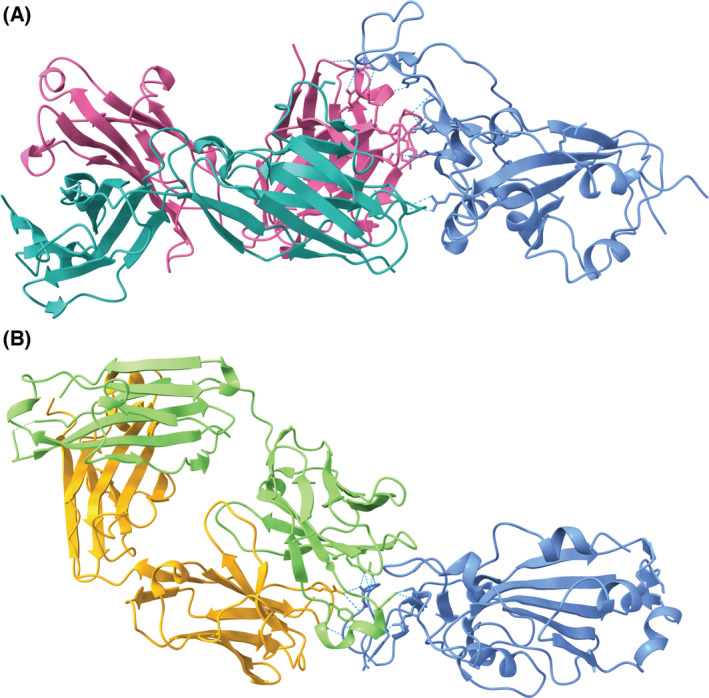
Fig. 4.

(A) Etesevimab (heavy chain: pink, light chain: teal) [40] binds to an epitope on a portion of the RBD of the SARS‐CoV‐2 S protein (blue) overlapping the ACE2 binding site (PDB ID: 7C01, visualized with ucsf chimerax, version 1.4) [41]. (B) Bamlanivimab (heavy chain: green, light chain: orange) [42] binds to an epitope on the RBD that overlaps with the binding site of etesevimab (PDB ID: 7KMG) [43]. Bamlanivimab is able to bind the epitope with the up and down conformations of its monomers [42]. Hydrogen bonds between each of the antibodies and the RBD are represented by dotted cyan lines.
