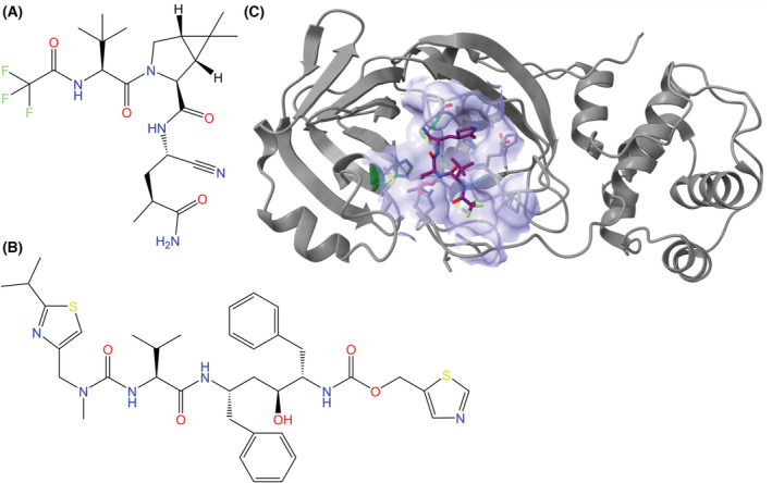
Fig. 9.

Chemical structures of (A) nirmatrelvir and (B) ritonavir, the two active components of Paxlovid. (C) Nirmatrelvir in complex with the Mpro of SARS‐CoV‐2 (PDB ID: 7SI9, visualized with ucsf chimerax, version 1.4) [89], with the binding pocket (lavender, composed of atoms within 5 Å of the inhibitor) visualized [82]. Nirmatrelvir is represented by a stick model with a purple carbon backbone. The peptide backbone of Mpro is represented in gray, with secondary structures displayed in cartoon format. In the binding pocket, stick models of amino acid residues are shown, and the catalytic dyad of Mpro (His41 and Cys145) is emphasized with carbon atoms in green. Hydrogen bonds between nirmatrelvir and Mpro are represented by dotted cyan lines, and heteroatom colors correspond to those in the 2D visualization.
