
Fig. 14.
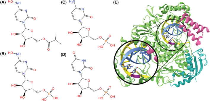
(A) Chemical structure of molnupiravir, an isopropylester prodrug of β‐d‐N4‐hydroxycytidine (NHC). (B) Chemical structure of NHC monophosphate. The active 5′‐triphosphate form of the drug is incorporated into the RdRp and induces viral activity. (C) Chemical structures of cytidine monophosphate and (D) uridine monophosphate, which NHC/Molnupiravir imitates. (E) RdRp complex (NSP12: green, NSP7: pink, NSP8: teal) bound to template (yellow) and primer (blue) RNA strands. NHC (purple backbone) is incorporated in the template strand and is base paired with adenosine (PDB ID: 7OZU, visualized with ucsf chimerax, version 1.4) [155, 156]. Hydrogen bonds between NHC and RdRp or the RNA strands are represented by dotted cyan lines, and heteroatom colors correspond to those in the 2D visualization.