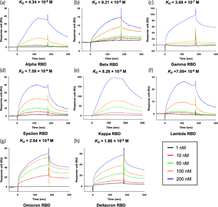
FIGURE 3.

SPR affinity analysis of the hCypA‐RBD variants. (a) [hCypA]‐[Alpha RBD], (b) [hCypA]‐[Beta RBD], (c) [hCypA]‐[Gamma RBD], (d) [hCypA]‐[Epsilon RBD], (e) [hCypA]‐[Kappa RBD], (f) [hCypA]‐[Lambda RBD], (g) [hCypA]‐[Omicron RBD], and (h) [hCypA]‐[Deltacron RBD]. The K D value of binding affinity is shown in Table 3.
