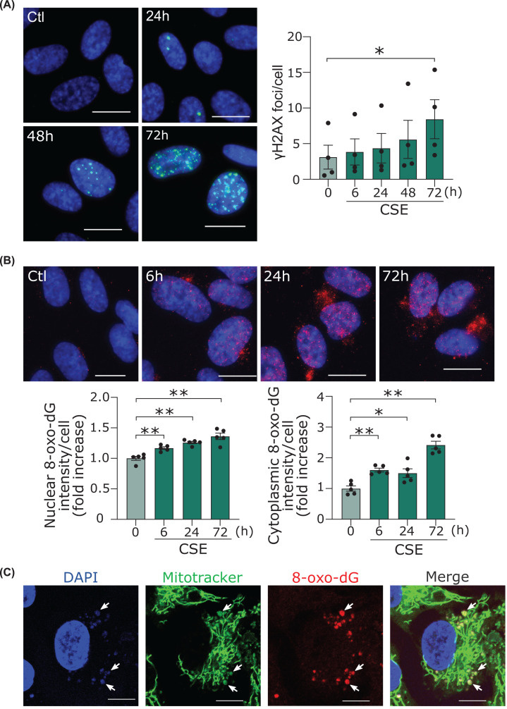
Figure 1. CSE increased nuclear and mitochondrial DNA damage in human endothelial cells.
(A) Immunofluorescent staining of the γH2AX (green) in HUVECs. Scale bar = 20 μm. Time course of γH2AX formation by CSE. *P<0.05 compared with corresponding control (n=4). (B) Immunofluorescent staining of the 8-OHdG (red) in HUVECs. Scale bar = 20 μm. Time course of the 8-OHdG formation in nuclei and cytoplasm. *P<0.05, **P<0.01 compared with control (n=5). (C) Images taken by confocal microscopy of immunofluorescent staining of the 8-OHdG (red) and MitoTracker™ RED CMXROS (green) and DAPI (blue) in HUVECs. Scale bar = 20 μm. Because permeabilization was not performed, the 8-OHdG in the nucleus was not stained. Arrows show colocalization of the 8-OHdG in mitochondria. Abbreviations: γH2AX, phosphorylated histone H2AX; 8-OHdG, 8-hydroxy-2′-deoxyguanosine; CSE, cigarette smoke extract.

