Figure 4.
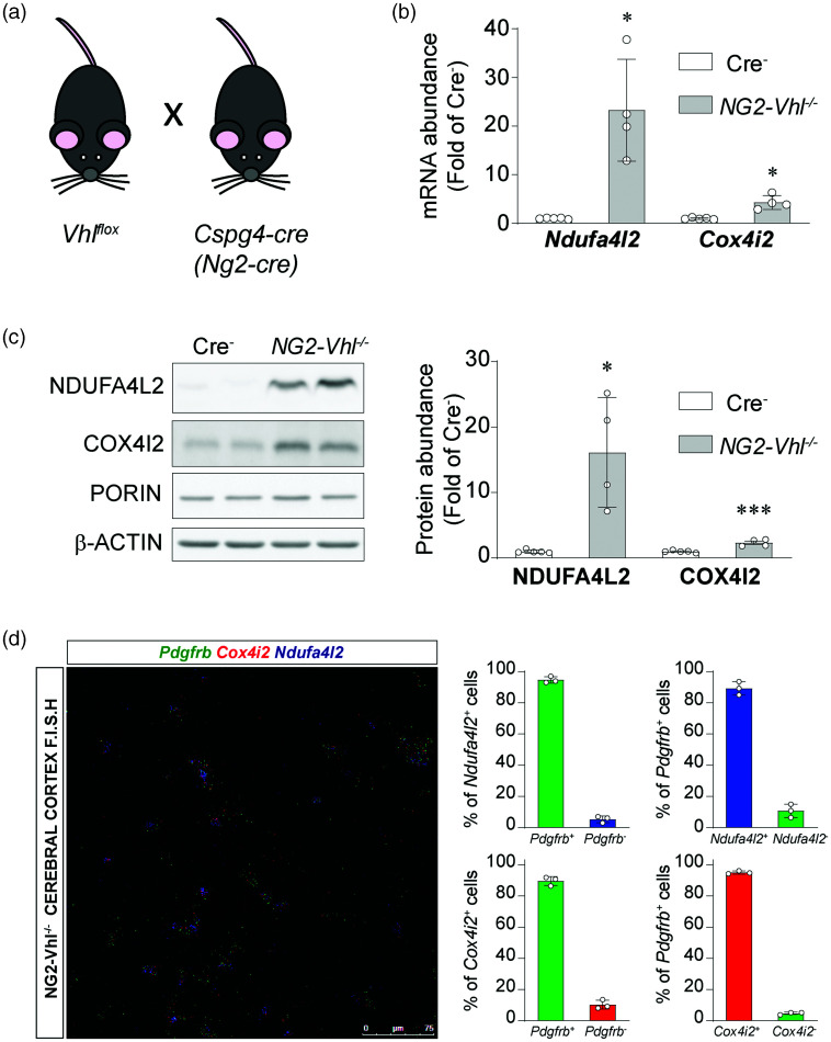
NDUFA4L2 and COX4I2 are induced in cerebral mural cells in NG2-Vhl−/− mice. (a) Graphical representation of breeding scheme. (b) Relative cortical mRNA transcript levels of Ndufa4l2 and Cox4i2 in homogenates from cortex of Cre− control and NG2-Vhl−/− mice (n = 4-5). (c) Representative western blot and quantification of NDUFA4L2 and COX4I2 expression in homogenates from cerebral cortex of Cre− control and NG2-Vhl−/− mice (n = 4-5) and (d) (Left) Representative image of multiplex RNA F.I.S.H. of NG2-Vhl−/− mouse cerebral cortex detecting Pdgfrb (green fluorescent signal), Cox4i2 (red fluorescent signal) and Ndufa4l2-expressing cells (blue fluorescent signal). (Right) Percentage of Pdgfrb+ and Pdgfrb− expressing Ndufa4l2 or Cox4i2 mRNA, and percentage of Ndufa4l2 or Cox4i2+ and Ndufa4l2 or Cox4i2− expressing Pdgfrb mRNA (n = 3) Data are represented as mean ± SD; Statistical analysis was performed using two-tailed Student’s t test with Welch’s correction when needed. *P < 0.05, ***P < 0.001, compared with Cre− control mice.

