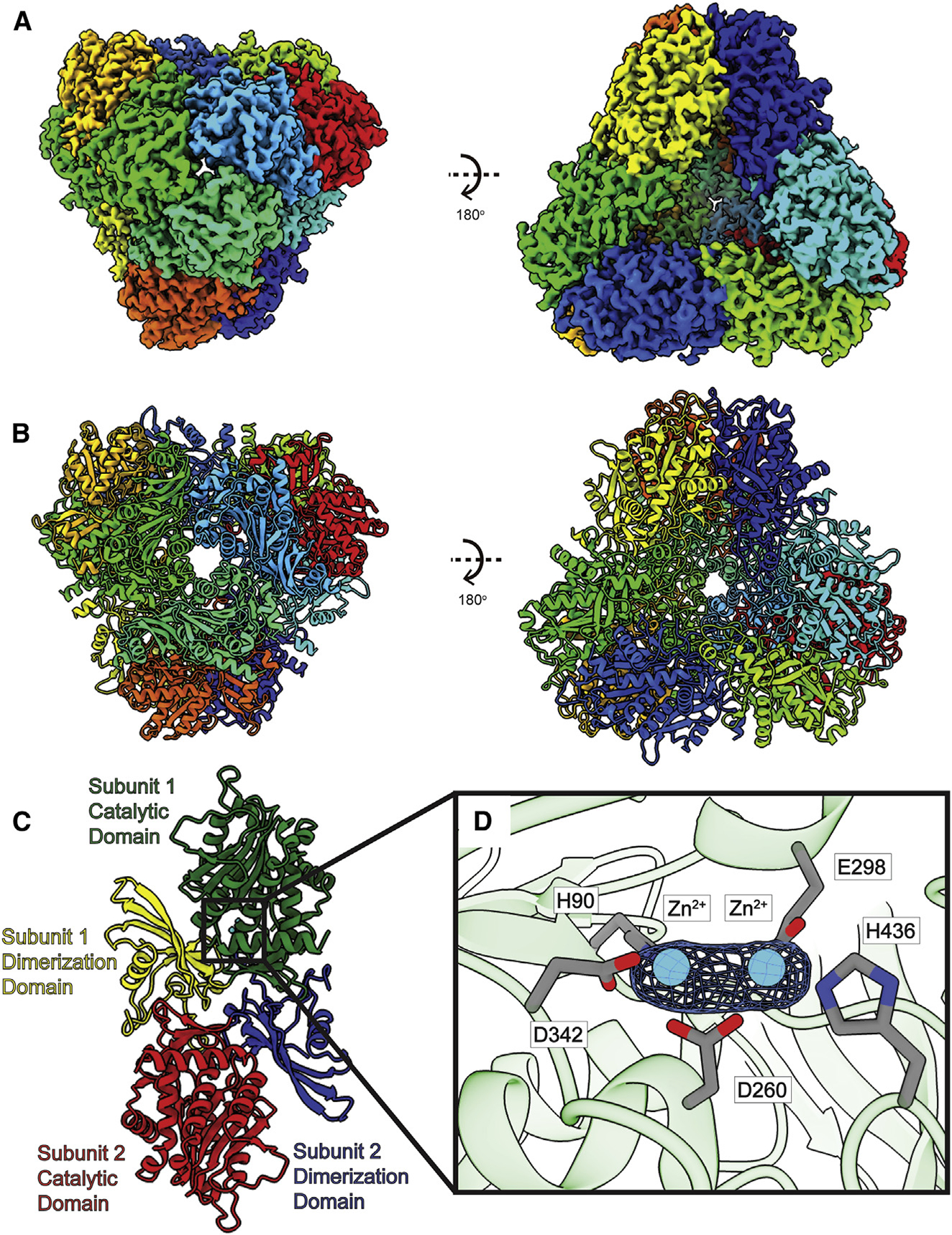
Figure 3. Structure of bovine DNPEP.

(A) Cryo-EM map of DNPEP.
(B) Structure of DNPEP. Twelve DNPEP monomers form a dodecamer with tetrahedral symmetry. Individual subunits are distinguished with different colors in both (A) and (B).
(C) Layout of DNPEP dimer. A DNPEP subunit can be divided into two units: a catalytic domain and a dimerization domain.
(D) Zoomed view of the Zn2+-binding site in a single DNPEP subunit. Two Zn2+ ions are found in each DNPEP subunit. The Zn2+ ions are colored cyan, cryo-EM densities (4σ) are depicted as blue meshes, and interacting residues are shown as gray spheres.
