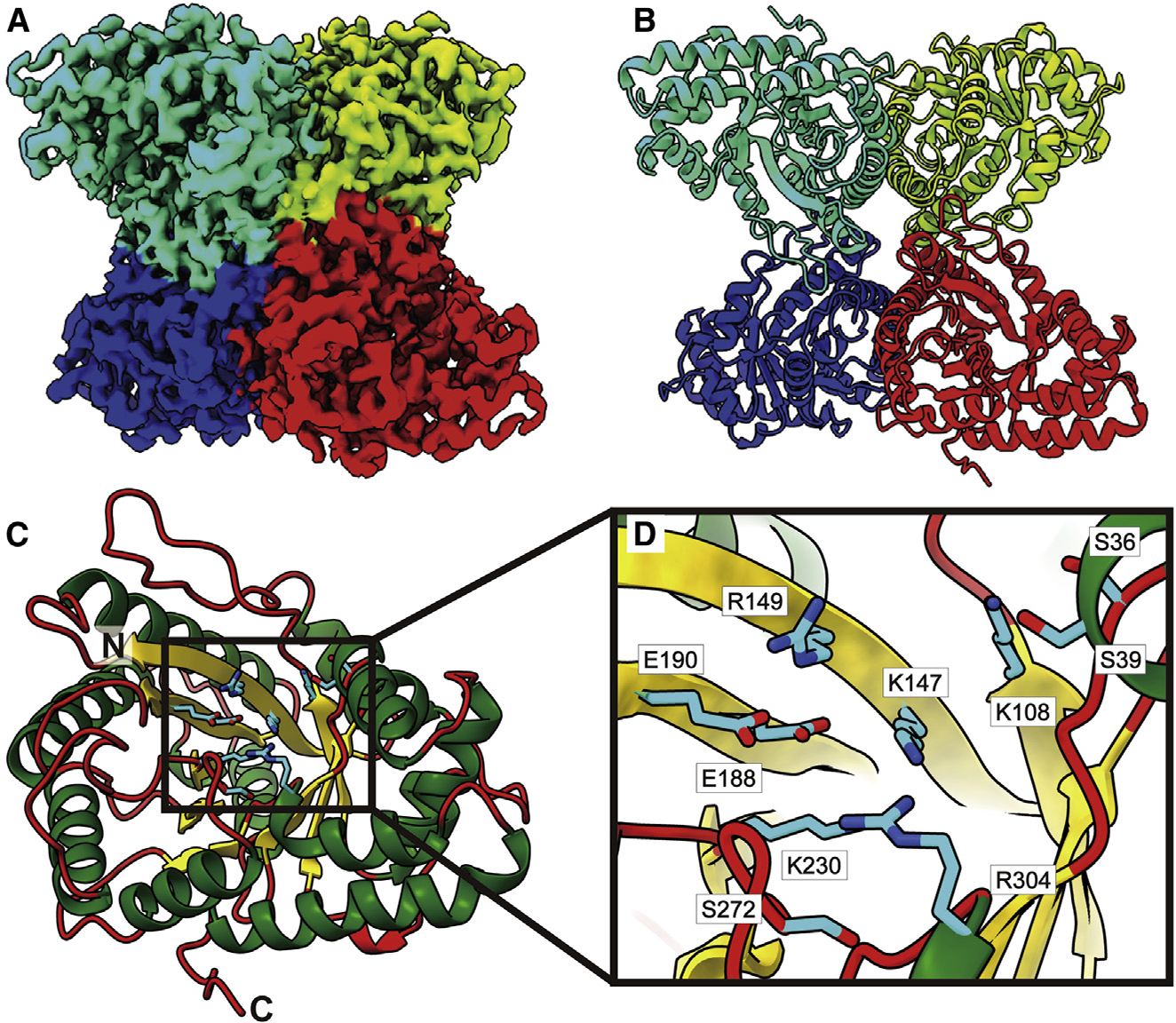
Figure 6. Structure of bovine FPA.

(A) Cryo-EM map of FPA.
(B) Structure of FPA. Individual FPA subunits aredistinguished by different colors.
(C) Structure of an FPA monomer.
(D) Zoomed view of the substrate-binding site. In both (C) and (D), the α helices, β strands, and flexible loops are colored green, yellow, and red, respectively. Residues forming the substrate-binding sites are in cyan sticks.
