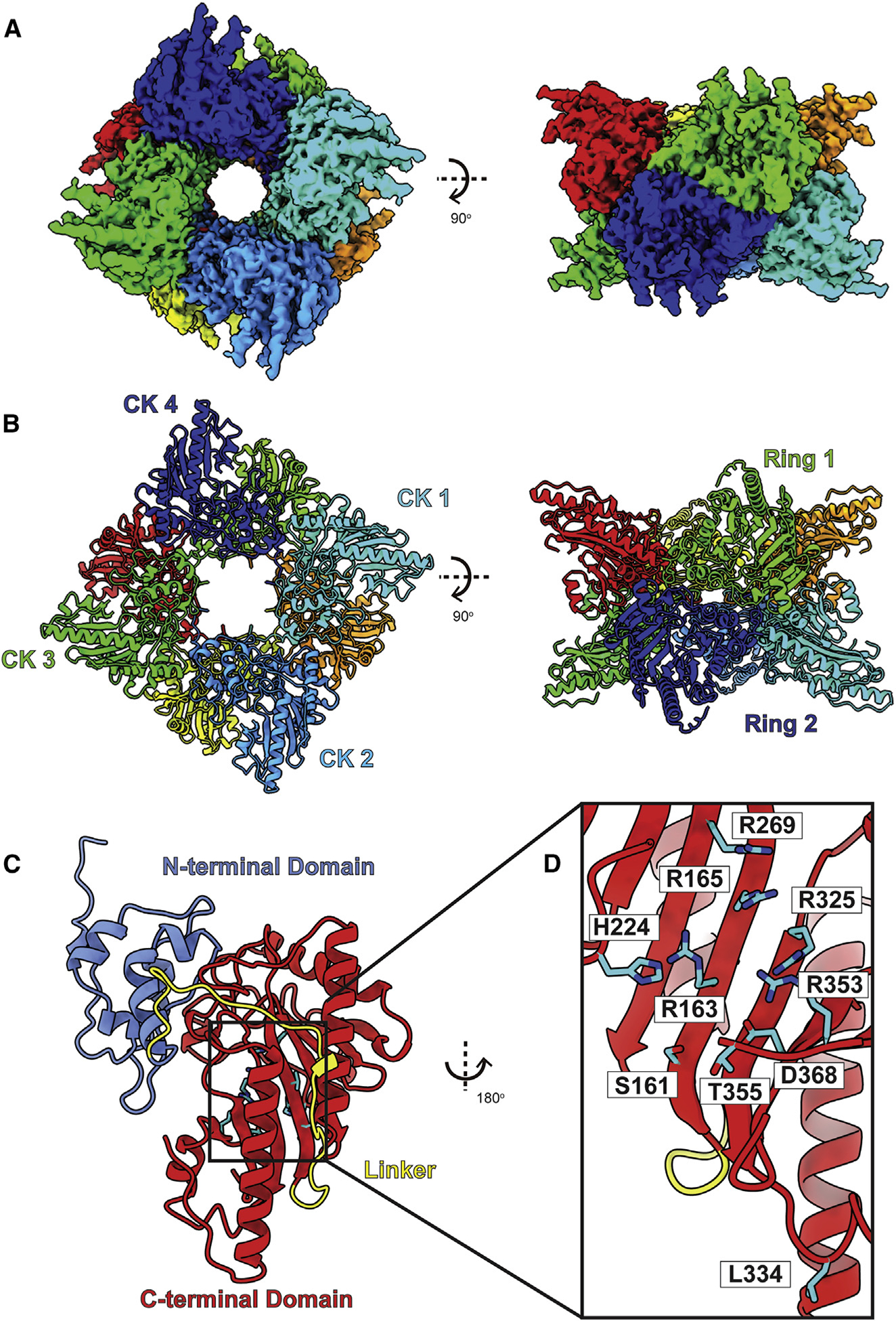
Figure 7. Structure of bovine uMtCK.

(A) Cryo-EM map of uMtCK.
(B) Structure of uMtCK. Individual uMtCK subunits are distinguished with different colors.
(C) Each bovine uMtCK subunit contains a small N-terminal dimerization domain (cyan) and a large C-terminal catalytic domain (red) connected by a flexible linker (yellow).
(D) Zoomed view of the substrate-binding site. Residues responsible for forming this site are in cyan sticks.
