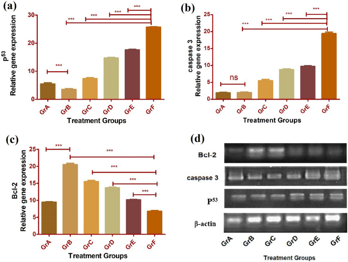
Fig. 8.
Comparative apoptotic gene expression analysis through RT-PCR with liver tissue samples from different experiential groups of animals (A-F). a Representing increases level of p53, b caspase-3, and c decreased level of Bcl-2 expression. Data indicated ± SD (n=3), (***) expressed significant (p < 0.05) upregulation or downregulation in gene, in conjugated nanoliposomes Apt-NLCs treated animals (GR F), samples in comparison to non-conjugated NLCs/ PEG-NLCs nanoliposomes (Gr D/F), free apigenin (Gr C) and carcinogen positive animals (Gr B). ns represented no significant changes among Gr A and Gr B for caspase expression assay. d Representing β- actin, p53, caspase -3, Bcl-2 gene expression of different experiential groups of animals in agarose gel electrophoresis study

