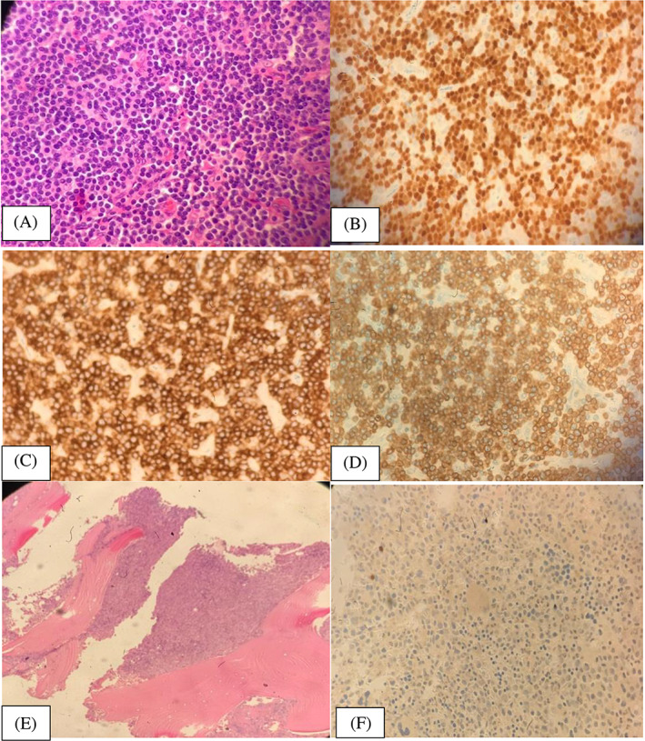
FIGURE 1.

(A) Lymph node histopathology, (B) lymph node immunohistochemistry for TDT, (C) is lymph node immunohistochemistry staining for CD7, (D) is lymph node immunohistochemistry for CD3, (E) shows a hypercellular marrow pre‐treatment and (F) is a negative TdT staining at diagnosis
