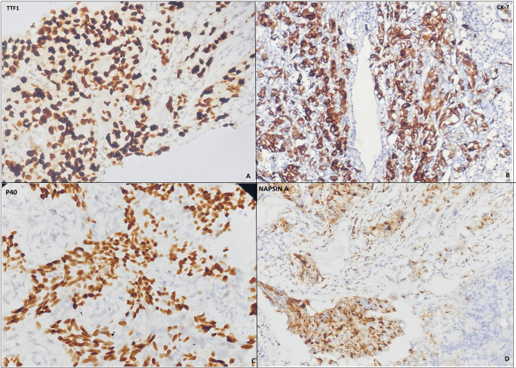
Figure 1. Photograph of IHC panel: (A) IHC of TTF1 shows the nuclear positivity in tumour cells; (b) CK-7 shows cytoplasmic staining in tumour cells; (C) P40 shows nuclear and cytoplasmic staining; (D) Napsin-A shows granular cytoplasmic and nuclear staining in tumour cells.
IHC: immunohistochemistry

